RELAST - development of a system for detecting reduced hypertrophy during scar formation
Project context
Within the collaborative project “Promotion of atrophic scar repair with elastin mRNA-based therapeutics”, a measurement system was developed for detecting reduced hypertrophy during scar formation. In clinical research, subjective scales are still often used to evaluate scars [1–3]. In recent years, the use of stereoscopic imaging techniques has been proposed [4]; however, previous approaches were either too inaccurate or not practical in real use.
Capture system
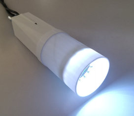
The goal was to develop a robust and practical system capable of capturing spatial structures of scar tissue. For this purpose, a camera system with an integrated lighting unit was developed that automatically illuminates the image field from four different directions and captures one image in each case. The capture cycle takes less than one second and enables the acquisition of parameters that correlate with scar hypertrophy.
Machine vision and analysis
The basis for the calculations comes from the shape-from-shading method [5]. The method is based on the assumption of constant illumination from defined directions – this is ensured by the device design. In addition, a continuous surface is assumed, which is usually the case for scar tissue. More difficult is the assumption of constant albedo, i.e. a uniform reflection of light on the surface [6]. Since skin surfaces can be smooth and reflective, it was evaluated whether image-correction methods could enable consistent analysis.
The generated images are homogenized and serve as the basis for algorithms that quantify the skin structure and the hypertrophy of the scar tissue. This approach enables an objective assessment of the scar structure that can be compared with clinical data.
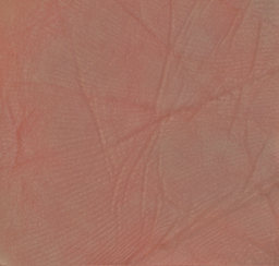
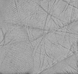 Skin image (homogenized) and the derived skin structure as the basis for quantification
3D visualization of the measurement results
The hardware-side development of the measurement system for detecting reduced hypertrophy was successfully completed. The system is available to clinical research under laboratory conditions to precisely measure scar structures and their conspicuousness. The functionality was demonstrated on sample specimens.
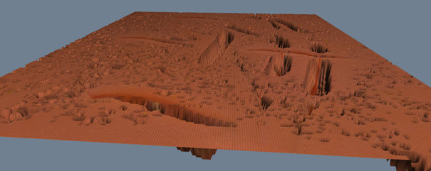 Example of the 3D visualization of the achieved measurement results
Download and funding
The full final report is available at: TIB – development of a measurement system for scars (final report)
Funded under the identifier E! 12338 RELAST by: Eurostars-2 joint programme with co-funding from the European Union Horizon 2020 research and innovation programme.


References
- Beausang E, Floyd H, Dunn KW, Orton CI, Ferguson MW. A new quantitative scale for clinical scar assessment. Plast Reconstr Surg. (1998) 102:1954–61.
- van de Kar AL, Corion LUM, Smeulders MJC, Draaijers LJ, van der Horst CMAM, van Zuijlen PPM. Reliable and feasible evaluation of linear scars by the patient and observer scar assessment scale. Plast Reconstr Surg. (2005) 116:514–22.
- Verhaegen PDHM, van der Wal MBA, Middelkoop E, van Zuijlen PPM. Objective scar assessment tools: a clinimetric appraisal. Plast Reconstr Surg. (2011) 127:1561–70. doi: 10.1097/PRS.0b013e31820a641a.
- Peake M, Pan K, Rotatori RM, Powell H, Fowler L, James L, Dale E. Incorporation of 3D stereophotogrammetry as a reliable method for assessing scar volume in standard clinical practice. Burns. 2019 Nov;45(7):1614–1620.
- Zhang R, Tsai P, Cryer J, Shah M. Shape from Shading: A Survey. IEEE Transactions on PAMI, 21(08), Aug 1999, pp. 690–706.
- Pentland AP. Local shading analysis. IEEE Transactions on Pattern Analysis and Machine Intelligence, 6:170–187, 1984.